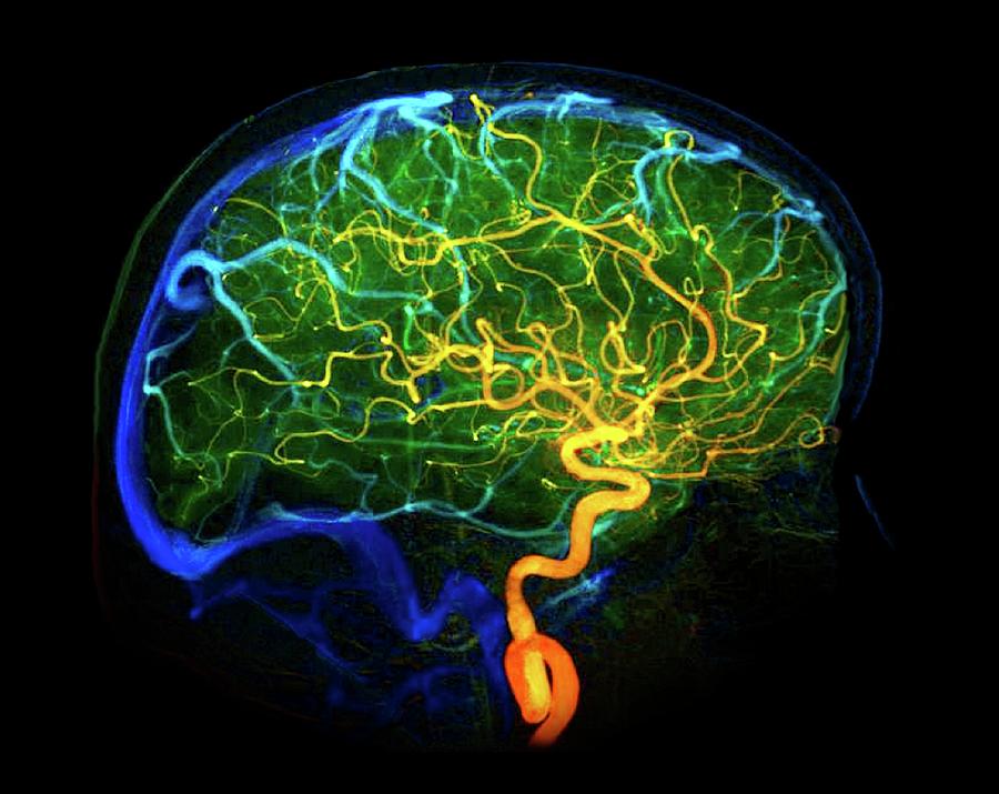
Blood Vessels Labeled Brain - Mri Blood Vessels In Brain Stock Photo - Download Image Now - iStock / It is a component of the cerebral circulation and is comprised of five arteries.. The major arteries supplying the brain provide the pia with its blood vessels. The space that separates the arachnoid and the pia is called the subarachnoid space. Arteries deliver oxygenated blood, glucose and other nutrients to the brain. These arteries arise in the neck, and ascend to the cranium. The vertebral arteries, and the internal carotid arteries.
The subclavian artery is divided into three parts based on anatomical landmarks. This can happen in either of two ways. This vessel supplies blood to the front part of your brain, knows as your frontal lobe. I) <rat brain vascular anatomy vasculature system of rat brain> yields ca. The brain blood supply functions
Brain Blood Vessels Photograph by Zephyr/science Photo Library from images.fineartamerica.com • the internal carotid artery passes through the temporal bone of the skull to supply oxygenated blood to the brain, eyes, forehead and part of the nose. The major arteries supplying the brain provide the pia with its blood vessels. The branches are differentiated into medial, mediolateral, and lateral portions, based on their localization and which anatomical territory they supply. This vessel supplies blood to the front part of your brain, knows as your frontal lobe. These include the common carotid artery that carries blood from the heart to the brain. The vertebral arteries, and the internal carotid arteries. Even if biology has never been your favorite subject, you still probably know a few basic things about the human body, including the fact that the heart pumps blood. Within the cranial vault, the terminal branches of these arteries form an anastomotic circle, called the circle of willis.
They run along either side of the neck and lead directly to the circle of willis.
Bulky middle tunic contains smooth muscle and elastin 3. The derivatives of the internal carotid arteries form the anterior blood supply (anterior circulation) of the brain, which includes the anterior and middle cerebral arteries. There are two paired arteries which are responsible for the blood supply to the brain; Label the blood vessels on the shin using the hints provided. The vessels that provide the organs with blood are called arteries. The subclavian artery is divided into three parts based on anatomical landmarks. The major arteries supplying the brain provide the pia with its blood vessels. I) <rat brain vascular anatomy vasculature system of rat brain> yields ca. This article lists a series of labeled imaging anatomy cases by system and modality. Cerebral circulation is the movement of blood through a network of cerebral arteries and veins supplying the brain. Learn vocabulary, terms, and more with flashcards, games, and other study tools. This resulted in fifteen trillion voxels, or individual. • the internal carotid artery passes through the temporal bone of the skull to supply oxygenated blood to the brain, eyes, forehead and part of the nose.
Start studying blood vessels of the brain. Blood vessels and the brain share a very important bond as blood vessels deliver oxygen and nutrients to all of the tissues and organs throughout the body, which is needed for the brain to function. Label the features of the heart using the hints provided. Several of the arteries of the circle of willis branch into smaller vessels that directly provide blood to the brain. Learn vocabulary, terms, and more with flashcards, games, and other study tools.
Vascular System Models - Arteries, Veins, Blood Cells - Complete Vessel Labeled Models | Cell ... from i.pinimg.com Within the cranial vault, the terminal branches of these arteries form an anastomotic circle, called the circle of willis. The subclavian artery is divided into three parts based on anatomical landmarks. If a stroke occurs in this area, you may see leg weakness and/or difficulty Blood vessel map reveals how brain gets food for thought. The external carotid arteries supply the face and scalp with blood. Branches arise from the circle to supply most of the cerebrum. The blood brain barrier keeps solutes from getting into the brain through blood vessels in addition, the blood brain barrier has cells which actually surround blood vessels on the outside with. The derivatives of the internal carotid arteries form the anterior blood supply (anterior circulation) of the brain, which includes the anterior and middle cerebral arteries.
Label the features of the heart using the hints provided.
This vessel supplies blood to the front part of your brain, knows as your frontal lobe. Ischemic strokes, which account for about 87% of all strokes, result when an artery that supplies blood to the brain becomes blocked by a clot. I) <rat brain vascular anatomy vasculature system of rat brain> yields ca. Veins take blood from cells and back to the heart and then to the lungs to be replenished with oxygen. The main arteries that supply the brain with blood are the paired vertebral and internal carotid arteries. The first part extends from its origin to the medial border of the scalenus anterior muscle. The pia, which covers the entire surface of the brain, follows the folds of the brain. Microscopically, it is formed by the endothelium of the blood vessel, capillary basal membrane, glial basal membrane and foot processes of glial cells. Each carotid artery branches into an internal. Label the blood vessels on the shin using the hints provided. Once in the cranial vault, the terminal branches form an anastomotic circle, commonly known as the circle of willis. Label the blood vessels of the posterior hip and thigh using the hints provided. Cerebral circulation is the movement of blood through a network of cerebral arteries and veins supplying the brain.
Willis' circle is located at the base of the brain, where it is joined by other vessels of the brain. The branches are differentiated into medial, mediolateral, and lateral portions, based on their localization and which anatomical territory they supply. The brain blood supply functions The major arteries supplying the brain provide the pia with its blood vessels. 3.860.000 results or ii) <rat brain vascular anatomy vasculature blood vessel system of rat brain> yields ca 399.000 results.
Circulatory Pathways | Anatomy and Physiology II from s3-us-west-2.amazonaws.com Use key choices to identify the blood vessel tunic described. Its smooth surface decreases resistance to blood flow They occur when a blood vessel in the brain bursts, spilling blood into the brain or the fluid that surrounds it. Each carotid artery branches into an internal. If a stroke occurs in this area, you may see leg weakness and/or difficulty Label the blood vessels of the posterior hip and thigh using the hints provided. The common carotid arteries have two divisions. It is a component of the cerebral circulation and is comprised of five arteries.
Branches arise from the circle to supply most of the cerebrum.
• the internal carotid artery passes through the temporal bone of the skull to supply oxygenated blood to the brain, eyes, forehead and part of the nose. This vessel supplies blood to the front part of your brain, knows as your frontal lobe. Cerebral circulation is the movement of blood through a network of cerebral arteries and veins supplying the brain. This resulted in fifteen trillion voxels, or individual. The blood brain barrier keeps solutes from getting into the brain through blood vessels in addition, the blood brain barrier has cells which actually surround blood vessels on the outside with. The first part extends from its origin to the medial border of the scalenus anterior muscle. Blood brain barrier blood brain barrier refers to the wall between the brain tissue and blood vessels. I) <rat brain vascular anatomy vasculature system of rat brain> yields ca. Blood is supplied to the brain, face, and scalp via two major sets of vessels: Scientists are developing new strategies for attaching drugs to molecules naturally transported across the barrier (labeled in green and blue). Several of the arteries of the circle of willis branch into smaller vessels that directly provide blood to the brain. The branches are differentiated into medial, mediolateral, and lateral portions, based on their localization and which anatomical territory they supply. It is a component of the cerebral circulation and is comprised of five arteries.
Blood vessel map reveals how brain gets food for thought blood vessels labeled. Microscopically, it is formed by the endothelium of the blood vessel, capillary basal membrane, glial basal membrane and foot processes of glial cells.
Blood Vessels Labeled Brain - Mri Blood Vessels In Brain Stock Photo - Download Image Now - iStock / It is a component of the cerebral circulation and is comprised of five arteries.. There are any Blood Vessels Labeled Brain - Mri Blood Vessels In Brain Stock Photo - Download Image Now - iStock / It is a component of the cerebral circulation and is comprised of five arteries. in here.